| |
|
|
| |
|
● Tissue engineering |
Lab on a chip research

Microfluidic Channel for Cellular Micropatterning and
Assay of Concentration-Gradient-Induced Migration |
| Objective&Results |
Recently, there has been considerable interest in the use of microfluidics
to miniaturize assays and control cellular microenvironments in cell-based
studies. We fabricated a microfluidic system in which cells attach only
at a central region along the microchannel, using which we assessed cellular
dynamic responses to a concentration gradient of biological factors (Fig.
1). In conventional methods, cells randomly attach to the entire inner
surface of the microchannel, making it difficult to precisely characterize
cellular responses and migration activities. Our system, however, consists
of three parts: a branched channel, a main channel for the cell culture,
and two side channels flowing into the main channel. The branched channel
is designed to generate a stable concentration gradient by mixing and dividing
two external inputs into six discrete streams. The main channel is coated
with a cell-repellent cross-linked albumin. A laminar flow of polyethyleneimine
(PEI) generated with the main and two side channels is used to change specific
regions in the main channel from being cell-repellent to cell-adhesive.
In this scheme, fibroblasts or hepatocytes are attached to the central
region along the main channel. Then the remaining surface is changed from
being cell-repellent to cell-adhesive with PEI, thereby facilitating cell
migration from the first site in response to a concentration gradient.
With this system, we have conducted cytotoxic assays with anticancer agents
and surfactants, as well as assays of migration across a concentration
gradient of a biological factor. Thus, this microfluidic system, combined
with our cell micropatterning technology, may be a useful tool in fabricating
controlled cell microenvironments for fundamental biological studies and
tissue engineering applications.

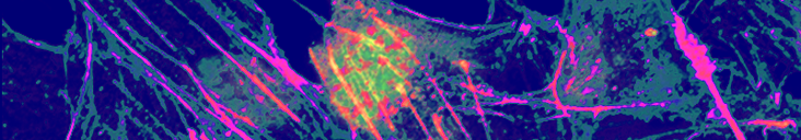
Fig. 1 Microfluidic cell culture device with concentration gradient generator.
|
| [Reference] |
T. Okuyama, H. Yamazoe, N. Mochizuki, A. Khademhosseini, H. Suzuki and
J. Fukuda*, Preparation of arrays of cell spheroids and spheroid-monolayer
cocultures within a microfluidic device, Journal of Bioscience and Bioengineering
(IF=1.71), 110, pp.572-6 (2010)
T. Okuyama, H. Yamazoe, Y. Seto, H. Suzuki and J. Fukuda*, Cell Micropatterning
inside a microchannel and assays under a stable concentration gradient,
Journal of Bioscience and Bioengineering (IF=1.71), 110, 2, pp.230?7 (2010) |
|
| ● Vascular |
| ● Liver |
| ● Hair |
| ● Pacnreas |
| ● Bone |
| ● Lab Chip/ MEMS |
| ● Surface modification |
| ● Microbe |
| |
| |
|
|
| |

|
|
 Fukuda Lab, Faculty of Engineering, Yokohama National University Fukuda Lab, Faculty of Engineering, Yokohama National University |
|