One of the major obstacles in engineering complex and thick tissue constructs is the requirement to fabricate vascular networks. Because oxygen is supplied only by diffusion, cells located more than a few hundred micrometers away from the surface of tissue constructs suffer from hypoxia and apoptosis. Therefore, a rapid fabrication strategy of spatially controlled capillaries is crucial for reconstructing three-dimensionally thick tissues. On the other hand, scaffold designed for tissue engineering have focused on modeling on extracellular matrix (ECM). In this study, we propose a technology for the detachment of cells from culture surfaces via an electrical stimulus, photocrosslinking scaffold and demonstrate that it is beneficial for the fabrication of vascular-like structures. |
We designed an oligopeptide, CGGGKEKEKEKGRGDSP, consisting of a cell adhesion
domain (RGD), alternate KE sequence, and cysteine (C). This oligopeptide
spontaneously binds to a gold surface via the gold-thiolate bond with C
and forms self-assembled monolayers with the electrostatic force of the
alternate positive and negative sequence (KEKEKEK). The gold-thiolate bond
could be reductively cleaved by applying −1.0 V vs Ag/AgCl reference electrode
and the monolayers were desorbed. Human umbilical vein endothelial cells
(HUVECs) were adhered to the gold surface via the RGD domain of the oligopeptide
and were then detached with the reductive desorption of the oligopeptide
by applying an electrical potential. We also designed photocrosslinkable
gelatin methacrylate hydrogels (GelMa) as ECM, which was gelled within
90 sec by light exposure (365 nm, 6.9 mW/cm2). By combining the electrical
cell detachment and the photocrosslinkable hydrogel, cells could be transferred
from a gold substrate to the hydrogel in a rapid and precise manner. The
procedures for the fabrication of capillary-like structures with this cell
transfer technology are presented in Fig. 1. Thin gold rods (600 µm in
diameter) were prepared by sputter-coating of Cr and Au layers on a glass
substrate. HUVECs were adhered on the rods via the oligopeptide and grown
to reach confluence for 3–4 days. The gold rods with cells were fixed in
a culture chamber, and 1.0 mL of the GelMa solution was then poured into
the chamber. After the gelation of GelMa by UV irradiation for 90 sec,
the rods were carefully extracted by applying a potential for 5 min Then,
the chamber was connected to a microsyringe pump, and the culture medium
including VEGF and FBS was perfused. In the coculture experiments, human
hepatoblastoma cells (Hep G2) were previously mixed in the GelMa solution
and poured into the chamber to fabricate liver-like tissue constructs.
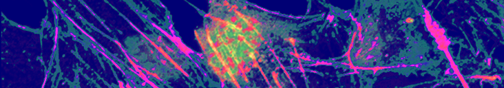
During the perfusion culture, the vascular-like structures were maintained
for at least 3 weeks. In the cocultures experiments, Hep G2 cells grew
in the hydrogel between the capillary-like structures (Fig. 2). According
to the image analysis, the number of Hep G2 cells was 10-fold greater than
the number of inoculated cells, indicating that this fabrication process
of the capillary-like structures is rapid enough to prevent oxygen depletion
and oxygen were supplied to Hep G2 cells through the HUVEC layers during
the perfusion culture.

Fig. 1 Scheme for the fabrication of the capillary-like structures in GelMa by using the electrical detachment of cells from the gold rods.

Fig. 2 Capillary-like structures in the hydrogel. Coculture of HUVECs (green) and Hep G2 (red). After 5 day
|